 TEKZARO
Enterprise
Meet a mouse whose brain cortex is made up of human cells
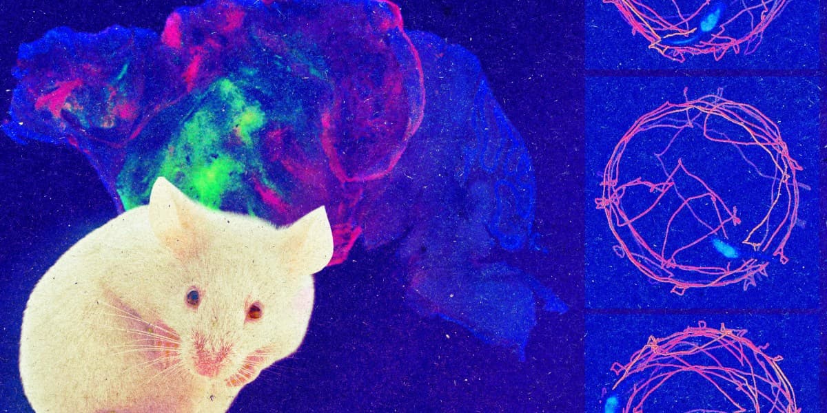
Stanford researchers have created mice that lack almost their entire cerebral cortex, then filled the empty space with human brain tissue grown from stem cells.
The resulting animals — the team calls them xenocortical mice — carry human-derived neural tissue that integrates into the mouse nervous system, forms working circuits and generates coordinated electrical activity. The study was published Wednesday in Nature.
Emptying the Cortex First
The technical move that makes this different from previous work is what the researchers removed before they added anything.
Human organoids have been transplanted into rodent brains before, and it works reasonably well. The problem is competition. As Sergiu Pașca, professor of psychiatry and behavioral sciences at Stanford and the study's senior investigator, put it, human cells get outcompeted by mouse cells very quickly — and it becomes difficult to disentangle the effect of the human tissue from the mouse tissue surrounding it.
So the team engineered the space away. They deleted ESCO2, a protein involved in cell division, specifically from cells expressing a marker for the dorsal and medial pallium — the embryonic forebrain regions that give rise to the cortex and hippocampus. The mice were also made immunocompromised so they would not reject human cells.
The result: animals missing 98 percent of their cortex and hippocampus, with roughly half the total brain tissue of a normal mouse.
They Were Surprisingly Fine
This is the detail that will stop most readers, and it stopped the researchers too.
Mice missing 98 percent of their cortex showed only minor behavioural changes — a more cautious gait was the main one. "These animals are not perfectly normal but also surprisingly functional, much more than we thought they would be," Pașca said.
It is a useful reminder of how much mouse behaviour runs on subcortical structures, and how far a rodent brain is from a human one in where its capabilities live.
What the Transplant Did
Human cortical organoids were successfully transplanted into 25 of 29 newborn engineered mice.
The tissue proliferated into the available space, growing 4.7-fold between two and three months after transplantation. The grafts integrated into the mouse nervous system, and the mice performed comparably to wild-type controls on motor, learning and social tests.
The grafts contained a diverse range of human cortical cell types, including von Economo neurons — rare cells found in large-brained social animals, implicated in several neuropsychiatric conditions, and notoriously difficult to produce in culture.
Calcium imaging of graft-derived neurons showed large synchronous bursts every few minutes, correlating with mouth and facial movements. Electrophysiological signals matched those bursts and were detected across the electrode array, which the researchers read as evidence that the graft forms a functional neural network.
Why Build This
The immediate application was modelling hypoxia — oxygen deprivation in brain cells, which around birth can cause cerebral palsy and is associated with autism and epilepsy. It affects humans considerably more severely than it affects mice, which is exactly the kind of species difference this model exists to capture.
The team exposed xenocortical mice, cortex-less mice and controls to low oxygen for five hours. Only the xenocortical mice were affected, showing trouble with balance and gait.
Hongjun Song of the University of Pennsylvania, who was not involved in the work, described the value plainly: a live model where these things can be tested with human cells in living animals.
Pașca sees two directions. Disease modelling — inducing a genetic mutation in control cells or transplanting a patient's own cells — and basic questions about what makes human and primate circuits distinctive, and how they process information early in development.
The Limitations Are Substantial
Several researchers were careful to bound the claim, and so were the authors.
Joseph Gleeson of UC San Diego put the constraint sharply. "It doesn't build a new cortex. What it does is it gives a lot of room for these organoids, or the cells in the organoids, to divide and grow and take up residence," he said, calling that both the single biggest finding and the single biggest limitation.
The grafted neurons are not organised into layers, which is a defining feature of actual cortex. The organoids lack inhibitory neurons entirely. Human neurons keep developing on their own slower timeline, so the model covers only the earliest stages of human development and is capped by the mouse lifespan. Variability in how individual organoids grow and integrate may limit which questions can be answered.
In-Hyun Park of Yale, also not involved, called replacing a substantial portion of an animal brain with human neural tissue an impressive technical advance — which is a precise compliment rather than a sweeping one.
Gleeson's summary was that this is about the best that can be done at this point.
The Ethics Were Built In
The team published details of the experiment's ethical oversight alongside the results, describing consultations with bioethicists and other neuroscientists throughout the project. Stanford separately convened a group of legal scholars, patient advocates, ethicists and scientists to oversee the work and provide feedback.
That is not a formality. As organoid research has advanced, the field has been working through questions about what sophistication threshold, if any, should trigger additional scrutiny — and a mouse whose cortex is made of human cells is the sort of result that makes those questions concrete.
Publishing the oversight process alongside the science is a reasonable norm for work in this territory, and it is worth noting that the researchers chose to do it rather than being made to.
Sources
Related Articles